Our team is using novel imaging techniques to give patients faster, more detailed diagnoses — all in a single scan
At Paul Strickland Scanner Centre, innovation is taking imaging to the next level. For years, MRI scans have been celebrated for their ability to visualise soft tissues with unparalleled clarity. However, when it comes to bones, traditional wisdom has always dictated: “Use a CT scan.” Now, thanks to our pioneering work using Deep Resolve software, that’s beginning to change.
Why Bones Matter in MRI
Bones have always been a challenge for MRI. The imaging relies on hydrogen atoms to generate its stunning visuals, and bone—dense and rigid—has very little free hydrogen to offer. As a result, bone structures typically appear black on MRI scans, providing little useful information. Yet, this is precisely the kind of data clinicians need to assess how cancers and other conditions are affecting skeletal structures.
Previously, doctors often relied on MRI to infer what might be happening around the bones but turned to CT scans to understand bone health itself. This meant scheduling additional appointments, creating delays in treatment planning. Our team at Paul Strickland Scanner Centre is changing that paradigm by harnessing the power of Deep Resolve, a deep learning-enhanced imaging technique.
A new vision with Deep Resolve
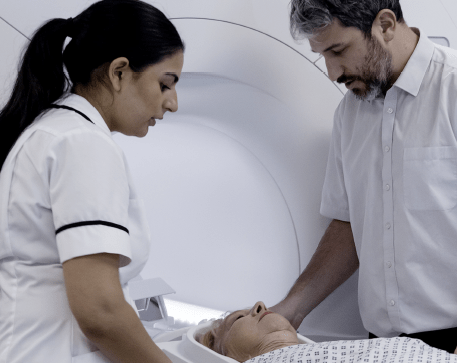
Deep Resolve leverages AI to enhance noisy, low-resolution images into something rich with detail. By refining how MRI captures and processes data, this approach allows radiographers to visualise bone structures alongside the soft tissues in a single scan. According to Will McGuire, a senior radiographer at the centre, “It’s a novel way of using the MRI scanner in a non-conventional way, addressing one of its major limitations. ”The results have been ground-breaking.” Will shares an example of a patient with renal cancer that had metastasised to the bones. Traditional CT imaging showed disrupted bone structures, but with Deep Resolve-enhanced MRI, the same abnormalities were clearly visible. This technique not only provides structural details but also integrates maps of cellular density, offering clinicians a comprehensive view of the disease’s impact.
‘This technique could revolutionise imaging’
Speeding up the process
Efficiency is another triumph of Deep Resolve. While older MRI techniques could take five minutes to scan a single section of the pelvis, newer iterations complete the task in about 30 seconds—a pace rivalling CT scans. “We’re fine-tuning the balance between scan time and resolution to ensure images are sharp enough for clinical use,” Will explains.
A new frontier
This capability isn’t just about convenience; it has real-world implications for treatment. For instance, in prostate cancer, distinguishing whether bone lesions are merely present or actively destructive can guide critical decisions about therapy.
Similarly, clinicians treating breast or kidney cancers now have a tool to assess bone involvement without needing additional scans. While the work is still in its early stages, the potential is immense. Will and his team are taking their findings to the prestigious European Congress of Radiology this year to showcase how this technology could revolutionise imaging worldwide. For the team at Paul Strickland Scanner Centre, this innovation reflects a commitment to pushing boundaries.
“We’re using the MRI scanner in ways it’s not traditionally been thought of,” Will says. And in doing so, they’re ensuring patients get faster, more detailed diagnoses—all in a single scan.