PET-CT scan
Information for Health Professionals
Everything you need to know about our PET-CT service at Mount Vernon Hospital and The Lister Hospital in Stevenage.

PET-CT scanning overview
Paul Strickland Scanner Centre first opened its doors in 1985, installing the UK’s first PET camera outside of central London, in 1991. In 2004, the Centre was able to upgrade to its first full PET-CT scanner, made by General Electric. A Siemens PET-CT scanner was also introduced in 2013.
In 2024, as part of a major refurbishment, the GE and Siemens scanners were decommissioned and a brand-new Siemens Biograph Vision 600 was installed. The introduction of the Biograph Vision 600 from Siemens Healthineers has revolutionised our imaging services. A vast upgrade on its predecessors, this high-quality scanner delivers clearer, sharper images whilst reducing scan times. This significantly benefits patients, ensuring we can keep pace with increasing referrals while maintaining the high standards.
The clinicians at the Centre pride themselves as being versatile and forward thinking, providing the highest standard of PET-CT service. Paul Strickland Scanner Centre is proud to hold UKAS BW 70000 Imaging Services Accreditation. UKAS assesses imaging services to ensure that the standard’s requirements are maintained through regular monitoring. Accreditation to standards is supported by NHS England, NHS Northern Ireland and NHS Wales, and recognised by the Care Quality Commission (CQC).
The Centre has several key advantages for people who need a PET-CT scan:
• As an independent charity it is buffered to a much greater extent from the current financial constraints of the NHS. This means that we are able to respond rapidly to advances in technology.
• Our PET-CT service runs 8am to 6.30pm Mondays to Fridays and occasionally we run weekend lists. This increases choice for patients, reduces the time patients have to wait before their scan, and reduces the time from initial presentation to diagnosis.
• Most radiopharmaceuticals we use are made at an on-site cyclotron. We are committed to serving our NHS patients and work closely with the Mount Vernon Cancer Centre (MVCC) and associated multi-disciplinary teams (MDTs). Clinicians often contact us to discuss urgent issues regarding diagnosis and treatment with our acclaimed, friendly and knowledgeable radiologists.
In 2018, we launched a satellite PET-CT site at Lister Hospital in Stevenage, offering improved access to patients living to the north of Mount Vernon.
Our team is research active and participates in clinical research trials.
Why choose us
We have been providing a high-quality service to clinicians and patients ever since Dr Paul Strickland launched the first PET imaging service at Mount Vernon in the 1990s, at the time the first service of its kind outside central London. With sites at both Mount Vernon and The Lister Hospital in Stevenage, we are able to accommodate patients from a wide catchment area.
About the team
We have a close-knit, experienced PET-CT team, consisting of booking administrators, radiography assistants, and radiographers/technologists. We also have on-site medical physics experts and radiologists devoted and dedicated to working together to provide a compassionate patient focused PET-CT service of the highest quality. All staff undergo regular continuous professional development (CPD) training to ensure they are aware of the latest developments in their areas of expertise and in compliance with regulatory requirements.
The service is overseen by our Lead Consultant for PET-CT, Dr Tony Chambers.
Dr Chambers graduated from Imperial College School of Medicine and is a Consultant Oncological and Radionuclide Radiologist at Paul Strickland Scanner Centre, Mount Vernon Cancer Centre and Lead for Nuclear Medicine and Uro-radiology at the London Northwest Healthcare NHS Trust. He is an ARSAC holder.
Dr Chambers has a long experience in PET-CT imaging for oncological and non-cancer indications, including dementia imaging. He has been an auditor for the national PET-CT reporting audit.
Our Superintendent Radiographer for PET-CT is Suzannah Patel.
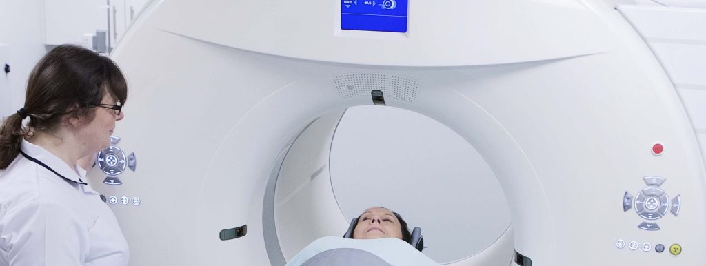
Our PET-CT scanner
The Siemens Biograph Vision 600 at Mount Vernon is used for whole body imaging, using radioactive glucose, i.e. 18F-Fluorodeoxyglucose (FDG), in people with cancer. Beyond FDG, PET radiopharmaceuticals specific for prostate cancer are used routinely; for example we are one of a select few centres in the UK who offer Prostate Specific Membrane Antigen (PSMA) PET-CT scanning for prostate cancer.
In addition, the centre provides a PET-CT service for appropriately investigating people who do not have cancer, including those with fever of unknown origin (PUO) and suspected vasculitis.
The centre’s PET-CT scanner has specific features that enable us to scan with low radiation doses, improving patient safety. Wide fields of view scanners, use of automatic dose reduction hardware and software techniques enable dose reduction while maintaining image quality.
PSMA PET-CT scanning
In February 2018, Paul Strickland Scanner Centre launched PSMA PET-CT scanning. The new scan could be particularly useful for men who have been treated for prostate cancer and are in remission (but under surveillance with regular blood tests) to make sure that they are clear of the disease. Some doctors believe that especially the relatively small number of patients who are at higher risk of developing metastatic disease (where the cancer spreads to outside the prostate gland) could really benefit, and it could mean catching and treating their cancer earlier – possibly resulting in a cure.
Example scan images
18F-PSMA PET-CT imaging is utilised in prostate cancer imaging
Areas of increased PSMA uptake on the PET component are mapped with a low dose CT scan to determine the extent of disease.
This study is highly sensitive and can identify sites of active nodal or metastatic disease that may not be visible on standard CT or MRI imaging alone.
This case demonstrates multiple sites of PSMA uptake within the bones that are not visibly abnormal on the CT component, these are in keeping with multiple deposits in the skeleton and will help the oncologists decide on next treatment options.

Click to enlarge
18-F FDG PET-CT Lung Cancer Case:
Identifying sites of disease not visible on conventional CT imaging (“Upstaging”)
The known site of lung cancer showed increased FDG uptake, as expected.
Increased FDG uptake was also identified in lymph nodes in the chest and in several bones (red arrows).
The PET-CT scan therefore demonstrated widespread metastatic disease that was not visible on the standard CT imaging.
This helped the Oncologists to chose a more appropriate treatment for widespread disease (i.e. chemotherapy) than the originally planned focal radiotherapy that would target only the primary cancer in the lung.

Click to enlarge