People with cancer are now able to benefit from lower radiation doses than ever before when having CT scans as part of their routine medical care and follow up. Dr Andrew Gogbashian explains.
People with cancer are now able to benefit from lower radiation doses than ever before when having CT scans as part of their routine medical care and follow up. Dr Andrew Gogbashian explains.
CT scanning has been well established in cancer care, for many years. CT scans are used for diagnosis. They are also used to measure whether treatment has started to work and to check whether a cancer has come back after treatment.
CT scans and X-rays both use radiation to produce a picture. As doctors use scans more and more, it becomes increasingly important to make sure patients are exposed to as little radiation as possible. The total dose a patient receives over the course of their lifetime is important, as well as the dose of each scan or X-ray. Of course, cancer specialists know that they need to keep this total radiation dose as low as possible. But they need to balance this with the benefits of scanning for each patient at various points in their treatment and after care.
The latest machines
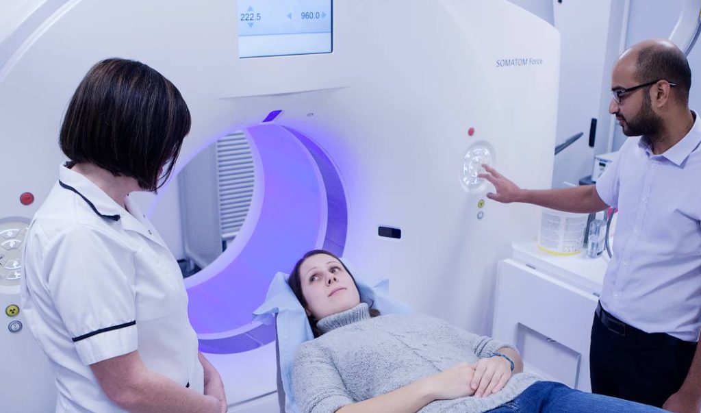
The Paul Strickland Scanner Centre has one of the most up-to-date CT scanners available – the Siemens Force. Our expert radiologists are able to reduce radiation doses while still producing excellent quality scans. The “Force” has several advanced features that can lower the radiation dose for each scan by between 30 and 70% compared to the average scan dose obtained with older scanning machines.
- “CARE dose 4D” automatically adapts the electrical current used to generate the X-rays to the shape and size of the patient, the part of the body being scanned and the angle of the scanner during the scan. Combining these makes sure that the lowest dose of X-ray radiation is used throughout the scan.
- “X-care” reduces the radiation dose for the most sensitive areas of the body, such as the breast, thyroid and eyes. It does this by automatically reducing the strength of the radiation beam when the patient is scanned from the front.
- “Care-KV” works alongside ‘CARE dose 4D’ to make sure the electrical voltage is adjusted along with the current. Both the current and the voltage that are used are then best suited to the patient and the aim of the scan.
- “Tin filter” blocks low energy photons, which aren’t needed for the scan. This directly reduces the radiation dose and also helps to make the scan clearer.
Improved software
CT scanners need to use computer software to turn the data they produce into pictures of good enough quality to be used medically. A process called ‘iterative reconstruction’ produces a series of images, each one a little clearer than the last until the best possible image is obtained. Using iterative reconstruction means that clearer scan images can be produced using lower radiation doses, but the drawback is that it’s a slow process. So it isn’t always practical when scans are needed without delay. Siemens ‘ADMIRE’ software (Advanced Modeled Iterative Reconstruction) can do this more quickly by using higher processing power.
Body tissues vary in how well they show up on a scan. Bones are dense, so are very clear. Air, water, areas of swelling and dead tissue are less dense, so don’t show up so well. This is important in cancer care as tumours often contain areas of less dense tissue. The ADMIRE software is better at picking out these low density areas from background ‘noise’ (graininess that affects the clarity of the scan). So clearer scans can be produced more quickly than ever before.
Patient benefit
Of course, patient safety is paramount at all ages, but the needs of younger patients has driven the development of newer and improved CT scanners that benefit everyone. Reducing radiation dose is particularly important for younger patients for two reasons. Younger body tissue can be at greater risk of radiation damage. And younger patients may need to be exposed to radiation from X-rays and scans later in life.
Testicular cancer provides a good example. These – generally younger – patients need to have between 5 and 10 follow up scans after their treatment, to make sure their cancer hasn’t come back. With an extremely high cure rate and hopefully a long life ahead of them, it’s particularly important to reduce their radiation exposure as much as possible. All treatment centres use radiation doses that are well inside the accepted safety range for their patients. But by utilising the Care-KV feature of our scanner, we are able to lower the radiation dose even further, while producing scans as clear as we got with our previous machines. Even without Care-KV, the other advanced features of the scanner mean that doses are still lower than they were.
When healthy people are having screening, it’s important to keep radiation exposure as low as possible. Lung cancer screening is now beginning to roll out in the UK. The Paul Strickland Scanner Centre is able to produce chest scans at extremely low dose – equivalent to the dose of two regular chest X-rays – making it ideal for this purpose.
The advanced features on our advanced CT scanner give us the opportunity to tailor scans to best suit each individual patient’s needs.