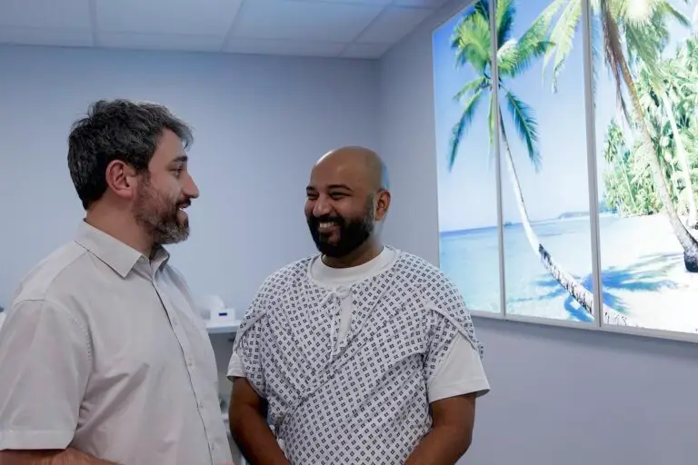
The UK is at the forefront of whole body MRI scanning worldwide. We have more experience than anywhere else in the world, having carried out more than 10,000 of these investigations. Dr. Anwar Padhani, Professor of Radiology at the Paul Strickland Scanner Centre, explains.
The UK is at the forefront of whole body MRI scanning worldwide. The Paul Strickland Scanning Centre has more experience than anywhere else in the world, having carried out more than 10,000 of these investigations. Dr. Anwar Padhani, Professor of Radiology at the Paul Strickland Scanner Centre, explains.
Whole body MRI scanning is one of the newest technologies available to help diagnose and monitor myeloma. It can be used if you have suspected or diagnosed multiple myeloma, or earlier conditions that may develop into full blown myeloma. These are solitary plasmacytoma, MGUS (“monoclonal gammopathy of undetermined significance”) and smouldering myeloma.

This type of scanning is now recommended in a number of myeloma guidelines produced for doctors, including by the UK’s National Institute of Health and Care Excellence (NICE).
Helping diagnose myeloma
In myeloma, whole body MRI scans can help to show whether myeloma is only in one place (localised) or spread throughout the body (systemic). By enabling your specialist to check the whole body for signs of myeloma, this type of scan can also confirm whether you have a solitary plasmacytoma, or myeloma outside the bones in soft tissues of the body. Whole body MRI is more reliable than X-ray at finding myeloma in soft tissues. Your specialist can also use MRI to guide taking biopsy samples.
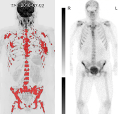
British Society of Haematology guidelines refer to a number of research studies showing that MRI is better than CT scanning at finding myeloma in the bones. MRI can detect up to 50% more myeloma lesions. Whole body low dose CT is an alternative, but is less sensitive than MRI at detecting when myeloma cells have invaded the bone marrow. MRI is also better at telling whether older patients have osteoporosis or degenerative disc disease, both of which can be easily confused with myeloma.
Whole body MRI is also useful when a patient has symptoms such as bone pain, but nothing shows on X-rays. By using specialist techniques, consultants in scanning (radiologists) can improve how well whole body MRI diagnoses smouldering myeloma or MGUS. These enhanced scans can pick up signs of myeloma cells in the bone marrow at an earlier stage – before they’ve had a chance to do any damage to the bone. These specialist MRI methods are called “diffusion weighted imaging” and “contrast enhancement”.
NICE guidelines, produced in 2016, argue that whole body MRI scanning saves money as well as improving patient health for patients with suspected myeloma. Because these scans are better at finding when smouldering myeloma or MGUS is developing into myeloma, doctors don’t need further tests before deciding on treatment. NICE say that whole body MRI should be the first choice of tests in patients suspected of developing myeloma. Whole body CT scan is another option, but NICE say it should only be offered if MRI is unavailable, unsuitable because of metal implants or refused by patients.
Monitoring myeloma
At the moment, doctors don’t routinely use whole body MRI to monitor treatment in myeloma patients, but it has a lot of potential. Specialist whole body MRI scans can show whether lesions in the bone marrow are active myeloma or scarring from previous treatment. This type of scan can also help myeloma specialists to monitor changes over time, showing how much treatment is helping or whether there are areas resistant to treatment. Although this approach isn’t yet fully established, The British Society of Haematology recommend its use where available.
In smouldering myeloma or MGUS, doctors know that you need to start treatment when myeloma cells start to multiply in the bone marrow or damage bone. The International Myeloma Working Group have produced guidelines for diagnosing when this is happening. They say that patients with smouldering myeloma should start treatment if their scan shows more than one area of myeloma in the bone marrow. Whole body MRI is more reliable at identifying these myeloma lesions at an earlier stage.
In more advanced multiple myeloma, MRI can help to show whether patients are at risk of complications, such as fracture or spinal cord compression. These patients can then have preventative surgery or radiotherapy, planned with the help of the scans. MRI can also distinguish between fractures caused by myeloma and non-cancer related fractures, which are common in older patients.
Prognosis
Understandably, patients often ask their doctors about their prognosis. Because it can pick up myeloma deposits that don’t show up on X-ray or CT, whole body MRI can help with this. Patients who have more than 7 lesions on whole body MRI generally have a poorer outlook, whereas patients with only one or two lesions have a better outlook.
Whole body MRI can reveal patterns of disease that provide information about prognosis. Myeloma can be concentrated in small, specific areas (focal disease) or spread more evenly through the bone marrow (diffuse disease). Diffuse disease generally indicates a higher risk of myeloma developing more quickly.
In smouldering myeloma or MGUS, whole body MRI is extremely important in providing information on prognosis. Any new findings on the scan indicate that myeloma is developing and treatment needs to start.
Follow up
MRI scanning isn’t currently recommended as routine follow up in MGUS or smouldering myeloma. But whole body MRI scans can help if levels of M protein (myeloma protein) in your blood start to go up, causing concern that the disease is becoming more active. Some patients have myeloma that doesn’t produce these marker proteins, or produces them in very low levels. In these cases, whole body MRI can be useful for monitoring how they are doing and is recommended in the British Society of Haematology guidelines.
Whole body MRI is also helpful in patients who have deposits of myeloma outside the bones.
The future picture
Using these advanced scanning technologies to help diagnosis and manage myeloma will change the likely outcome for the next generation of myeloma patients. It is now possible to detect active disease and start treatment at a much earlier stage, before there is any evidence of bone damage. We hope this will mean that people stay well for longer and with fewer complications from their myeloma.